What Is a Suture Anchor in Orthopedics?

What Is a Suture Anchor in Orthopedics? It is a small implant that fixes soft tissue to bone. Surgeons use it to repair tendons, ligaments, and labral tissue. Common sites include the shoulder, hip, knee, ankle, and foot. The device may be made from metal, PEEK, or bioabsorbable materials. It usually carries one or more strong sutures.

The need for these repairs is substantial. The World Health Organization reports that musculoskeletal conditions affect about 1.71 billion people worldwide. Its data also identifies low back pain, fractures, and osteoarthritis as major causes of disability. These figures do not measure suture-anchor use directly. Still, they show the wider clinical burden behind orthopedic reconstruction. Grand View Research’s orthopedic devices analysis also describes continued market growth, driven by sports injuries, aging populations, and surgical innovation.

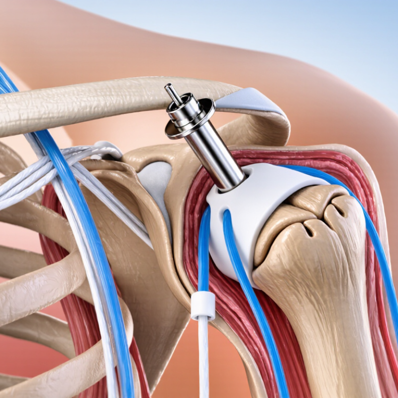
In Suture Anchor Orthopedics, fixation quality depends on more than the implant alone. Bone density, anchor design, insertion angle, suture strength, and rehabilitation all influence the result. Picture a small anchor seated inside the humeral head, holding a repaired tendon against prepared bone. That simple image hides complex biomechanics. Some anchors provide excellent fixation. Others may face pullout, breakage, migration, or inflammatory reactions.

Clinical judgment remains essential. Product brochures cannot replace peer-reviewed evidence or surgeon experience. Research methods also vary, making direct comparisons difficult. The FDA’s device records and published clinical studies can support safer evaluation. Patients should discuss implant material, expected healing, possible complications, and rehabilitation timelines with a qualified orthopedic specialist.
What Is a Suture Anchor in Orthopedics?

Definition and Role of Suture Anchors in Orthopedic Surgery

A suture anchor is a small orthopedic implant that attaches soft tissue to bone. It usually contains an anchor body and strong surgical sutures. Surgeons place it into prepared bone, then pass the sutures through a tendon, ligament, or labral tissue. This creates a stable point for repair during healing.

The anchor’s role is mechanical support. It holds the repaired tissue close to its original attachment site while new biological connections develop.

In shoulder surgery, it may secure a torn rotator cuff or labrum. In the ankle, it can help repair damaged ligaments. The surgeon selects the anchor type, size, and placement according to bone quality, tissue condition, and the repair pattern.

Placement requires careful control. Poor positioning can irritate nearby structures or weaken fixation. Excessive tension may also restrict natural tissue healing. The fit matters. In clinical practice, imaging, examination, and surgical experience guide decisions more reliably than a single implant feature. Patient age, activity level, smoking history, and rehabilitation habits can influence outcomes. An anchor supports repair, but it does not replace biological healing. Even with precise surgery, recovery may be uneven, and some repairs can fail.

This is where the explanation feels incomplete: surgical technique matters greatly, yet postoperative loading is often underestimated. Gradual rehabilitation, supervised movement, and honest reporting of pain remain essential parts of the repair process.


Main Components and Materials of a Suture Anchor

What Is a Suture Anchor in Orthopedics?

Main Components and Materials of a Suture Anchor

A suture anchor is a small implant that fixes soft tissue to bone. Surgeons commonly use it in shoulder, hip, knee, and foot repairs. Its main parts include the anchor body, eyelet, and attached suture. The eyelet guides the thread and reduces rubbing during knot tying. Some designs use knotless locking mechanisms. Others require secure knots placed beside the repair.

Anchor materials affect strength, handling, and tissue response. Common choices include titanium, stainless steel, PEEK, bioabsorbable polymers, and biocomposite materials. Titanium offers strong fixation but can create imaging artifacts. PEEK is radiolucent, which helps postoperative imaging. Bioabsorbable materials gradually break down, although degradation is not always predictable. A 2023 systematic review in Arthroscopy reported that anchor-related complications remained uncommon, but definitions varied between studies. That limitation deserves attention.

Tips: Match the anchor to bone quality, tear size, and loading demands. Check the eyelet before insertion. A damaged suture may fail before the anchor does. Grand View Research projects continued growth in the global suture anchors market through 2030, driven by sports injuries and minimally invasive repairs. Market growth does not prove clinical superiority. Selection still depends on anatomy, surgeon experience, and patient factors.

How Suture Anchors Are Placed and Secured in Bone

What Is a Suture Anchor in Orthopedics?

Suture anchors are small fixation devices used to reattach soft tissue to bone. Surgeons commonly use them for torn tendons, ligaments, and labral tissue. Placement begins after the surgeon examines the injury and prepares the surgical field. The damaged tissue is identified through an incision or camera-assisted approach. Bone preparation must be controlled. Excessive drilling can weaken the attachment site.

The surgeon creates a pilot hole at the planned repair location. Its depth and angle depend on the joint, bone quality, and anchor design. Some anchors require tapping, while others are inserted directly. After insertion, the anchor expands, screws in, or locks beneath the bone surface. The fit matters. Sutures attached to the anchor are then passed through the torn tissue. They may be tied with knots or secured using a knotless mechanism. Gentle tension brings the tissue back toward its original footprint.

The repair is checked through movement and visual inspection before closure. Poor tension can restrict motion, while insufficient fixation may reduce stability. Small errors can matter. In my clinical reading, bone quality often receives less attention than the anchor itself, although it strongly influences security. Not every anchor performs identically in every patient. Age, bone density, tear pattern, and healing capacity all affect decisions. Rehabilitation protects the repair while new tissue gradually bonds to bone. Healing takes time.

Common Orthopedic Procedures Using Suture Anchors

A suture anchor is a small implant that secures tendon, ligament, or labral tissue to bone. Surgeons place it through a tiny drill hole, then pass strong sutures through damaged tissue. The method can reduce incision size, but healing still depends on biology, rehabilitation, and patient behavior.

Common orthopedic procedures using suture anchors include arthroscopic rotator cuff repair, Bankart repair, SLAP repair, and hip labral repair. In shoulder surgery, anchors reattach torn tendon edges to the humeral head. In instability surgery, they fix the labrum around the socket. Hip surgeons may use them to restore the labrum and improve the joint seal. The American Academy of Orthopaedic Surgeons reports that about two million Americans seek medical care for rotator cuff problems each year. The 2024 American Joint Replacement Registry Annual Report also shows continued growth in monitored shoulder procedures, although registry data do not measure every anchor repair.

Tips: Ask whether the anchor is absorbable or permanent. Confirm the planned rehabilitation timeline. Do not judge success by pain alone. Strength often returns slowly.

Anchor choice depends on tissue quality, tear pattern, bone density, and surgical technique. A larger anchor is not automatically better. That assumption deserves scrutiny. Complications can include stiffness, infection, anchor loosening, or repeat tearing. AAOS clinical guidance emphasizes shared decision-making because imaging findings do not always match pain or function. Experienced surgeons combine examination, imaging, and patient goals before selecting an anchor-based repair.

What Is a Suture Anchor in Orthopedics?

A suture anchor is a small implant used to attach soft tissue, such as a tendon, ligament, or labrum, to bone. The chart shows representative midpoint values from commonly reported clinical ranges for the number of anchors typically used in selected orthopedic procedures.

Typical anchor numbers vary according to tear size, tissue quality, fixation technique, anatomy, and surgeon preference. The displayed values represent approximate midpoints of commonly used clinical ranges rather than fixed requirements.

Benefits, Risks, and Factors Affecting Anchor Selection

What Is a Suture Anchor in Orthopedics?

A suture anchor is a small fixation device placed in bone during orthopedic repair. It connects strong sutures to damaged tendons, ligaments, or labral tissue. Surgeons commonly use anchors in shoulder, hip, knee, and foot procedures. The sutures hold repaired tissue against bone while biological healing develops. In practice, anchor repair can support stable movement and smaller incisions than some traditional techniques. It may also reduce disruption to nearby tissue. However, smaller does not mean risk-free.

Possible complications include infection, nerve irritation, anchor loosening, bone damage, stiffness, and continued pain. Poor placement can weaken the repair or injure nearby structures. Healing also depends on blood supply, tissue quality, rehabilitation, and patient health. Anchor selection requires careful judgment. Surgeons consider bone density, repair location, tear size, expected loads, and the number of fixation points. They may also compare anchor size, insertion method, suture strength, and whether the material remains permanently or gradually absorbs. Patient activity matters too. A competitive athlete may place different demands on a repair than an older adult with fragile bone. Imaging and direct surgical findings can change the plan. No anchor works perfectly in every situation. That limitation deserves honest discussion before surgery. Recovery instructions remain essential, even when fixation appears secure.

What Is a Suture Anchor in Orthopedics? - Benefits, Risks, and Factors Affecting Anchor Selection

Category Key Information Clinical Relevance
Definition A suture anchor is a small implant used to attach soft tissue, such as a tendon, ligament, or labrum, to bone. It provides a fixation point while the repaired tissue heals and integrates with the bone.
Common Orthopedic Uses Rotator cuff repair, shoulder labral repair, elbow ligament repair, hip labral repair, ankle ligament repair, and selected foot and knee procedures. The indication depends on the injured structure, tissue quality, location, and required fixation strength.
Basic Components Most systems include an anchor body and one or more sutures or tapes. Some designs incorporate an eyelet or suture-passing mechanism. The design affects how the implant is inserted, how the suture is managed, and how tissue compression is achieved.
Common Materials Anchor bodies may be made from metallic alloys, nonabsorbable polymers, or bioabsorbable or biocomposite materials. Sutures are commonly made from high-strength synthetic polymers. Material choice influences imaging characteristics, mechanical behavior, degradation, and the potential for material-related reactions.
Insertion Methods Anchors may be inserted through a predrilled or tapped bone hole, or secured using an impact-driven mechanism, depending on the design. The technique must match the anchor design and account for bone density, hole size, insertion angle, and proximity to other structures.
Potential Benefits Enables soft-tissue-to-bone fixation, supports minimally invasive or arthroscopic procedures, may reduce the need for larger bone tunnels, and can allow multiple fixation points. These benefits may help restore anatomy while limiting surgical exposure, although outcomes depend on the overall repair and rehabilitation plan.
Mechanical Risks Possible problems include anchor pullout, loosening, breakage, suture rupture, tissue cut-through, malposition, and inadequate fixation. Risk is affected by load, insertion technique, bone quality, anchor placement, tissue quality, and patient adherence to postoperative restrictions.
Biological and Surgical Risks Infection, inflammation, foreign-body response, fluid-filled bone cavities, cartilage or tendon damage, stiffness, nerve or vessel injury, and the need for revision surgery may occur. The actual risk varies by procedure, patient health, implant material, surgical technique, and postoperative care.
Anchor Size Available sizes differ in diameter, length, and suture capacity. The anchor should fit the available bone and the intended repair configuration. An oversized implant may weaken or fracture bone, while an undersized implant may provide insufficient fixation.
Bone Quality Bone mineral density, cortical thickness, local anatomy, and the presence of cysts or previous tunnels can influence fixation strength. Poor or compromised bone may require a different anchor design, altered placement, augmentation, or another fixation strategy.
Tissue and Repair Requirements The surgeon considers tissue thickness, tear pattern, retraction, tension, the number of fixation points, and whether simple or sliding knots are needed. Suture configuration and anchor placement should distribute load without excessive tissue strangulation or gap formation.
Material Selection The choice between metallic, polymer, and bioabsorbable or biocomposite materials depends on the surgical site, imaging needs, degradation profile, and surgeon preference. No single material is ideal for every procedure; benefits and limitations should be assessed for the individual case.
Imaging Considerations Metallic anchors may create imaging artifact, whereas some polymer-based materials are less visible on certain imaging studies. Expected follow-up imaging and the need to evaluate recurrent symptoms may influence material selection.
Patient Factors Age, activity level, smoking, diabetes, bone health, immune status, medications, previous surgery, and ability to follow rehabilitation instructions are relevant. Patient-related factors can affect healing, infection risk, fixation durability, and the timing of return to activity.
Postoperative Care Protection of the repair, staged range-of-motion exercises, strengthening, wound monitoring, and gradual return to activity are commonly required. Premature loading can increase the risk of repair failure, while excessive immobilization may contribute to stiffness.
Key Selection Principle Anchor selection should balance fixation strength, bone preservation, tissue handling, material behavior, imaging requirements, and the surgeon’s familiarity with the technique. The most suitable anchor is procedure-specific and should be selected by the treating orthopedic surgeon after evaluating the patient and repair plan.

Medical information is provided for educational purposes only and does not replace personalized advice from a qualified orthopedic professional.